cardiac

Pulmonary Embolism [McConnell's Sign] | R Panjwani MD, A Lew MD, S Laymon MD, D Guha MD | Bronx NY

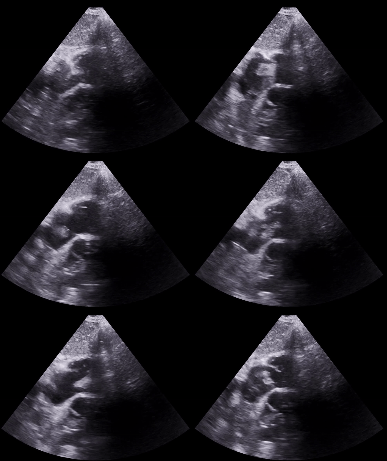
Clip 1: Apical 4 chamber view. Dilated right ventricle (RV) with an akinetic RV free wall and apical sparing consistent with McConnell’s sign.

By Dr. Reema Panjwani:

An elderly female patient with a history of heart failure (Ejection Fraction 45%--> 50% in 2022) and diabetes presented to the emergency department for a fall and syncope. She says she woke up in the morning feeling lightheaded in bed and fell, was concerned, and called an ambulance. She had been feeling short of breath for the last few days. Vital signs were notable for: blood pressure 85/50 mm Hg, a heart rate of 110 a minute, and saturating 90% on room air. The patient was alert and oriented, tachypneic, cool to touch, diaphoretic, and was answering questions very slowly. 

POCUS Echocardiogram apical 4 chamber view (clip 1 above) showed a dilated right ventricle, an akinetic RV free wall that spared the apex (ie, McConnell’s sign). The inferior vena cava (clip 2 below) was plethoric with less than 50% variation with respirations. These findings were suspicious for obstructive shock secondary to massive pulmonary embolism.

As CTA Pulmonary Embolism study was being completed, a saddle pulmonary embolus was identified on wet read. The Pulmonary Embolism Response team activated. As the patient was being transported back from the CT room to the resuscitation bay, she started to have worsening altered mental status. On two occasions, she lost palpable pulses requiring ACLS protocol to be initiated, including the administration of epinephrine and TPA. ROSC was achieved after 1-2 rounds of compression after each occurrence, requiring synchronized cardioversion for monomorphic ventricular tachycardia. A central line was placed, and the patient was started on three pressors, and a TPA infusion prior to being taken to the intensive care unit in preparation for mechanical thrombectomy. 

Reema’s takeaway: Always include pulmonary embolism in your differential for syncope. Subsequently, include the POCUS Echocardiogram as part of your workup in a syncopal patient.

After successful thrombectomy and placement of an IVC filter, the patient was discharged from the hospital a couple weeks later in good health.

Clip 2: Plethoric inferior vena cava, almost not change in diameter with respiration.