An elderly female is brought into your resuscitation bay feeling like it’s hard to breathe for almost a week, with bad dyspnea on exertion, and a nagging non productive cough at rest. She used to smoke years ago, picking it up in the 70s and quitting in the 90s. No history of heart or lung problems that she can recall. She’s clearly not in extremous, seemingly comfortable and answering your questions appropriately. Oxygen saturation is 92% on room air, with systolic blood pressure 140s mm/Hg. Heart rate in the low 100s. Oxygen saturation comes right up to 100% on 2L nasal cannula. Docs in the room report ‘audible wheezing’. A portable chest radiograph, image 1 below, is concerning for dense consolidation versus pleural effusion creating an almost complete white out appearance of the left lung. POCUS thoracic: shows a unilateral pleural effusion on the left (image 2 below), no consolidation. What’s going on here? When there’s fluid in the chest, the sound beams emanating from your transducer love the fluid medium, swim through it with ease, and light up the thoracic spine (image 2, positive ‘spine sign.’). So the fact that we can see the thoracic spine on the left means there’s fluid in the chest. Conversely, image 3 below of the right hemidiaphragm shows a negative ‘spine sign,’ meaning no pleural effusion on the right. Those same sound beams in a healthy right chest are encountering air, which does not transmit sound well at all. As a result, you can’t see the thoracic spine on the right. It’s still there, you just can’t see it.

Image 1: Single view portable chest radiograph shows white out of almost entire L lung.
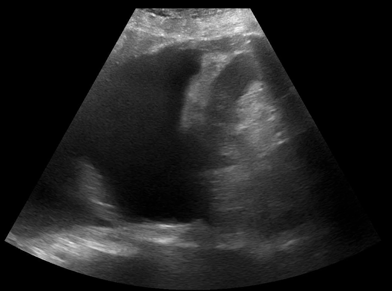
Image 2: POCUS thoracic of the left hemidiaphragm with a positive ‘spine sign.’
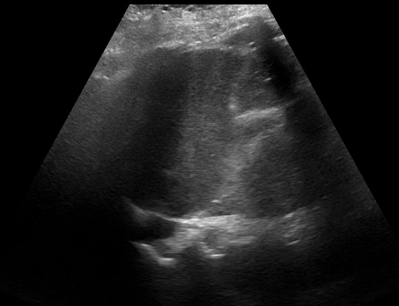
Image 3: POCUS thoracic of the right hemidiaphragm with a negative ‘spine sign.’