A 60s F is brought into your resuscitation bay by a prehospital crew with acute shortness of breath. She’s sitting bolt upright, gasping for air. Treatment prior to arrival includes albuterol nebulizer treatments and intramuscular epinephrine for asthma / COPD. She’s oxygenating in the low / mid 80s. Blood pressure is 180s/110s mmHg.
Not all that wheezes is asthma - so the classic medical aphorism goes.
POCUS cardiothoracic in the right hands quickly elucidates the real culprit - sympathetic crashing acute pulmonary edema. Nitroglycerin drip and positive pressure ventilation turns this situation around in minutes. A word to the wise, in addition to cardiothoracic POCUS, please do get your chest radiographs, brain natriuretic peptides, and lung auscultation in the acutely dyspneic patient. The dilemma, if I may, is when those things are done in lieu of cardiothoracic POCUS.
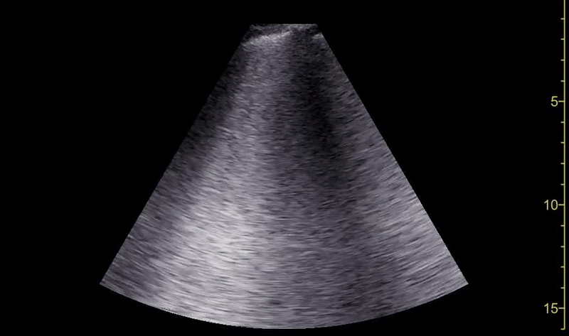
Image 1: Diffuse B lines left thorax.
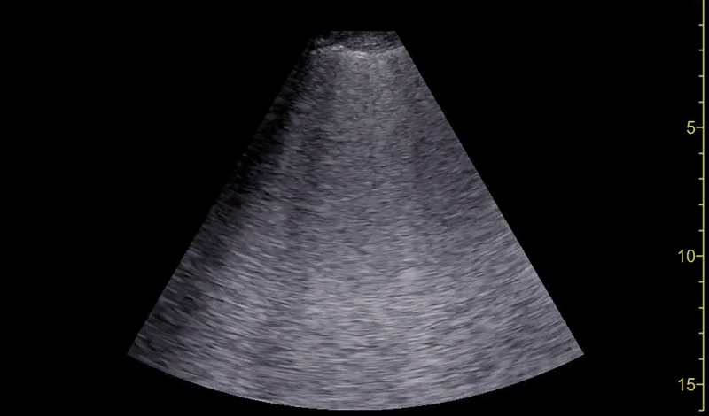
Image 2: Diffuse B lines right thorax.
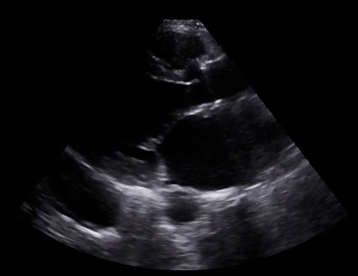
Image 3: Severely reduced systolic function.