A middle aged male police officer is carried into front triage by his colleagues. He’s shot in the lower left abdomen. In the resuscitation bay he’s clammy and diaphoretic, in shock, but awake. Blood Pressure 70/40 mm/hg, HR 80s, RR 18, 100% SP02. Large bore IV access, immediate blood products transfused. This patient is going to the operating room for an exploratory laparotomy, but is there also a problem in the chest? Love The Three Box Concept of the EFAST from colleagues out in California, USA, and nicely incorporated here. High suspicion for hemorrhage from the abdomen in this case, but did that bullet also ricochet up into the chest to cause a hemothorax/pneumothorax. Or did it traverse the mediastinum and cause tamponade? In 2026 a complete EFAST incorporates an eval of all three boxes, the abdomen, the thorax, and the heart.
EFAST: Box 1: The abdomen. There’s a very high suspicion for hemoperitoneum, so the team starts in the most sensitive anatomic location, the hepatorenal space, specifically the caudal tip of the liver. It’s positive for free fluid (Clip 1), so we’re done with the abdomen, zero reason to then evaluate the left upper quadrant or the pelvis. There's blood in the abdomen, we’re done with Box 1. If the hepatorenal space were negative, now we must then look at the lesser sensitivity zones of the left upper quadrant and the pelvis. Box 2: The thorax. There’s lung sliding and a negative spine sign, so no clinically significant pneumothrorax or hemothorax (Clips 2 and 3). Box 3: The heart. Any echo window will do, clip 4 is the parasternal long axis showing no pericardial effusion in between the pericardium and the descending thoracic aorta.
The patient did very well.
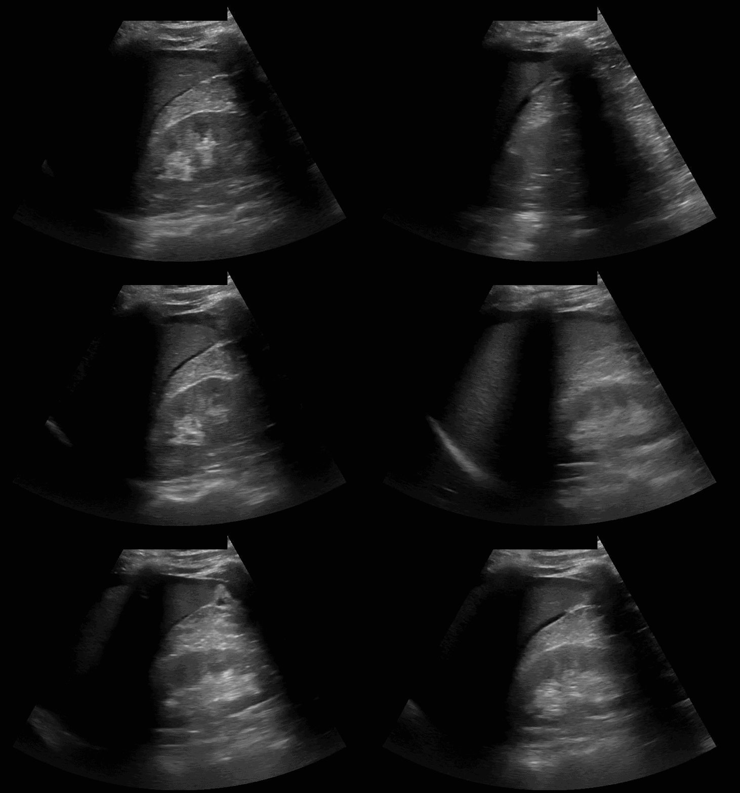
Clip 1: Free fluid in the hepatorenal space
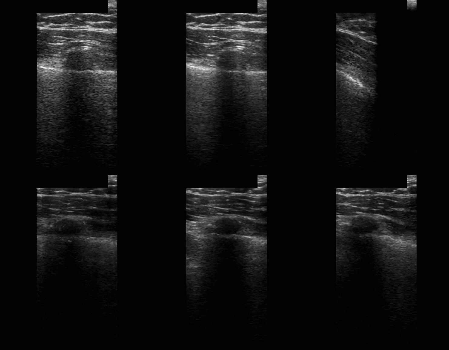
Clip 2: Lung sliding. With a linear transducer (most sensitive) in the anterior chest, lung sliding rules out a clinically significant pneumothorax.
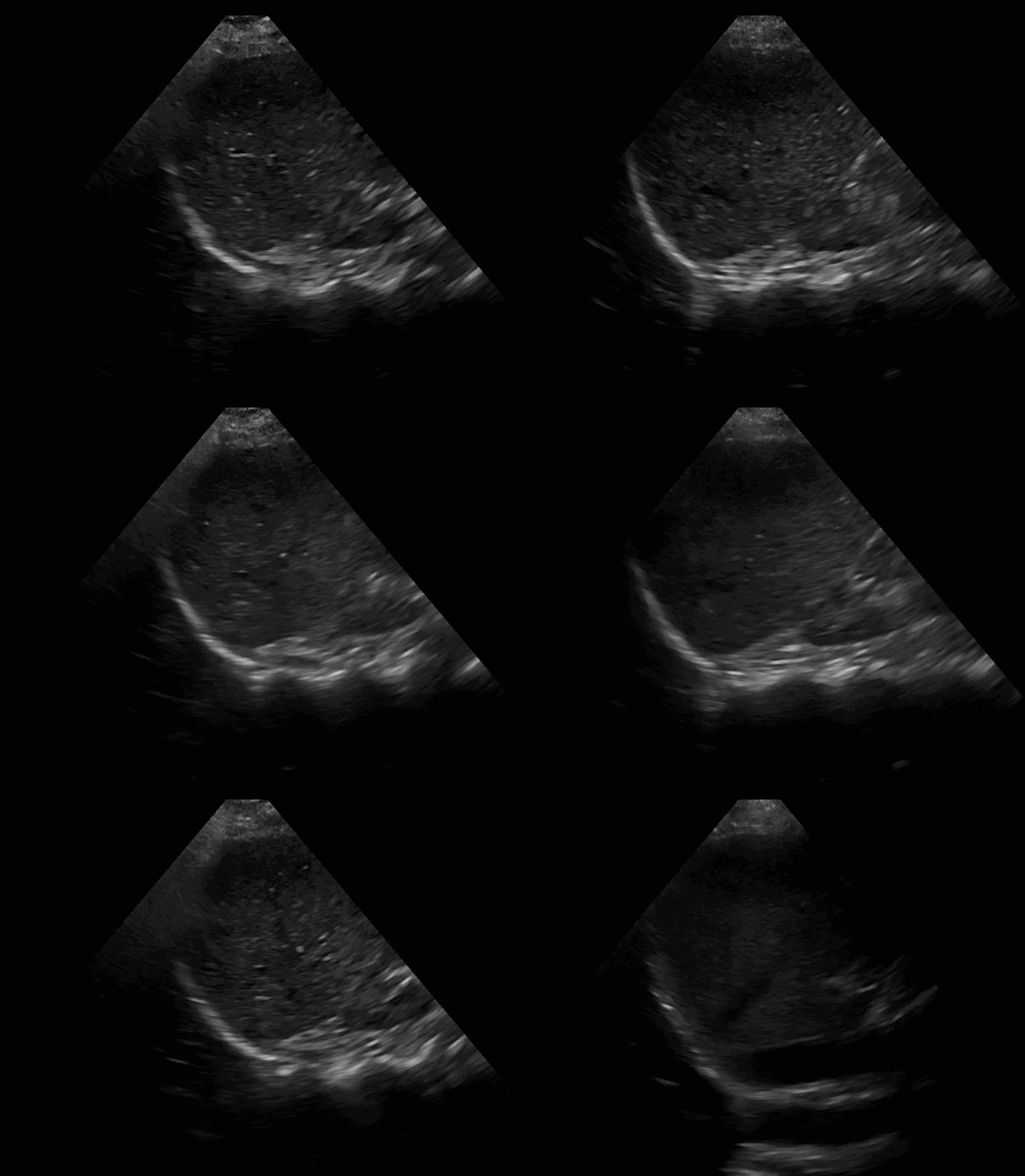
Clip 3: A negative spine sign, the intersection of the diaphragm and thoracic spine shows no hemothorax.
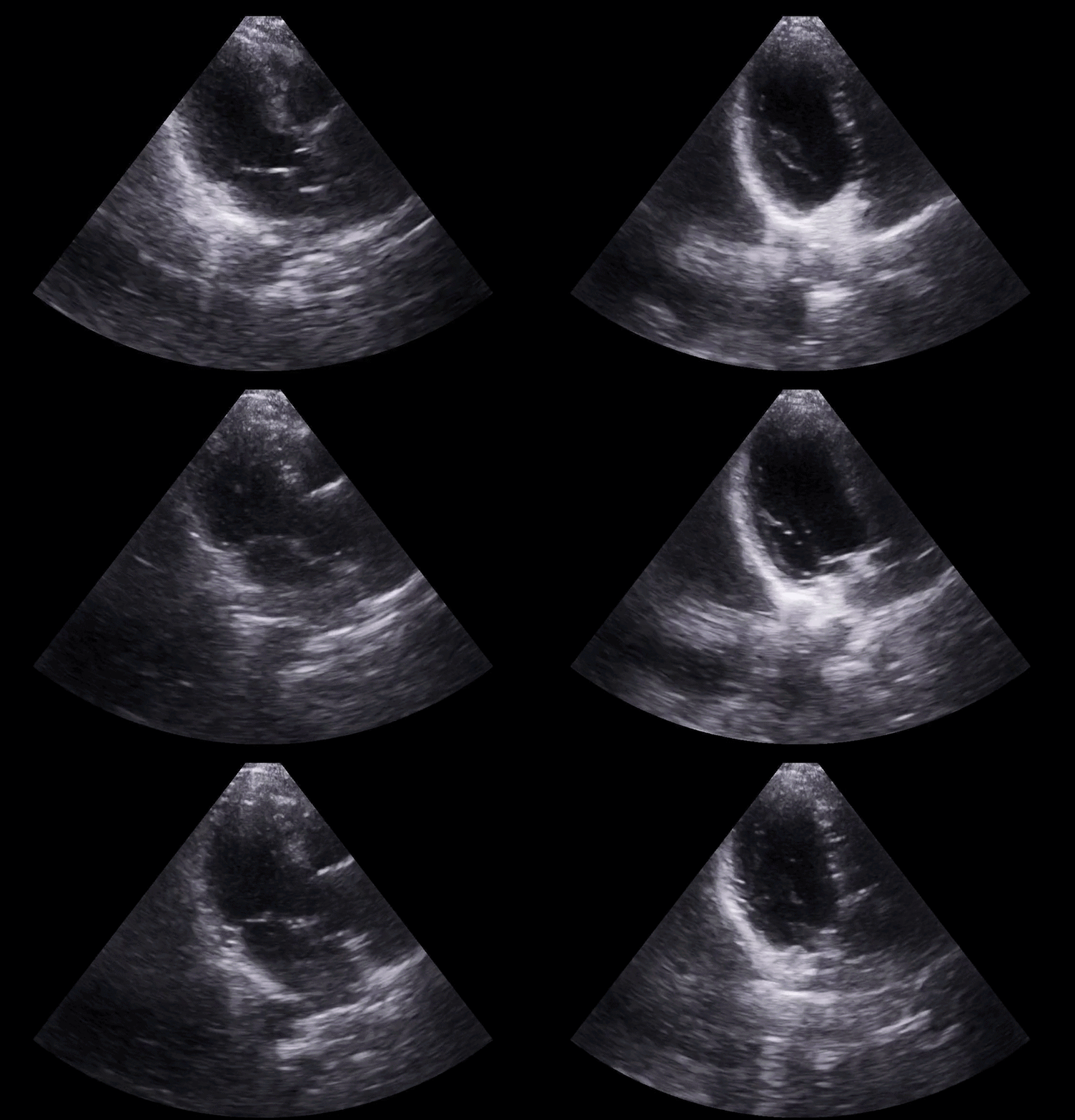
Clip 4: Parasternal long axis without pericardial effusion.