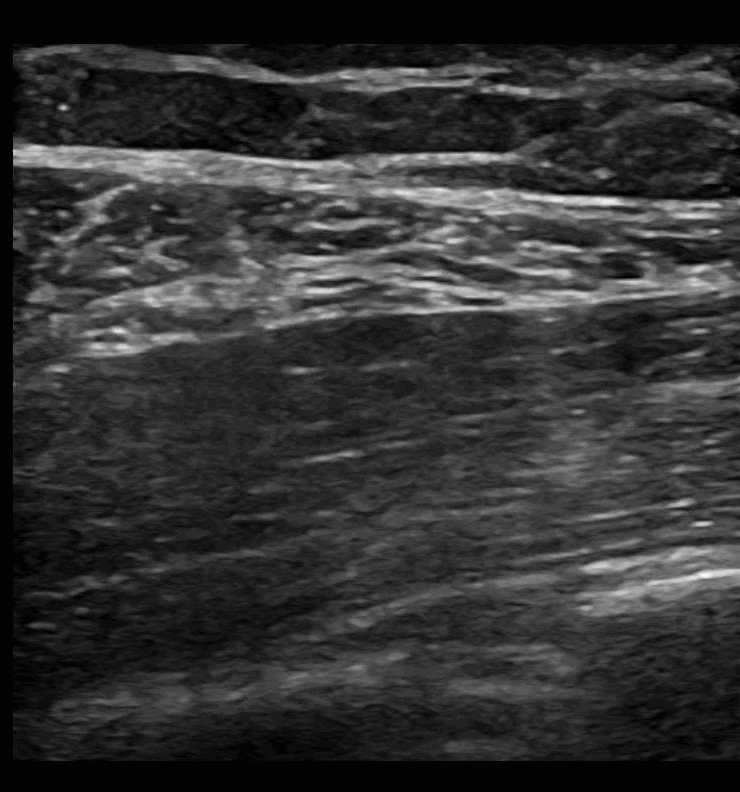
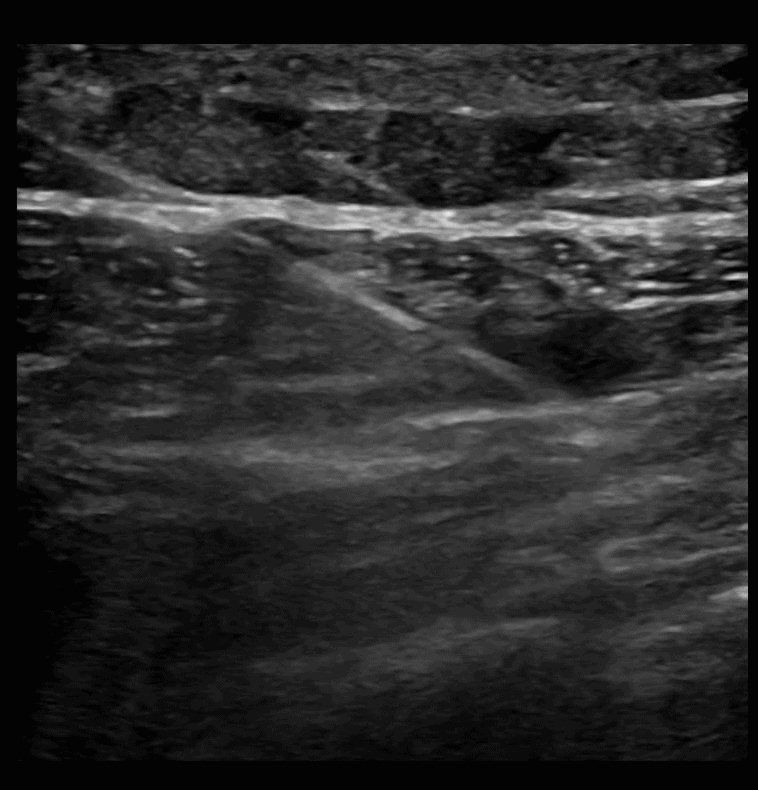
SAPB 1: Pre-scan. The latissimus dorsi muscle comes to a point on the right of the screen as you scan posterior to anterior. Just deep to the latissimus dorsi muscle is the serratus anterior muscle. Deep to the serratus anterior muscle is the rib and pleura.
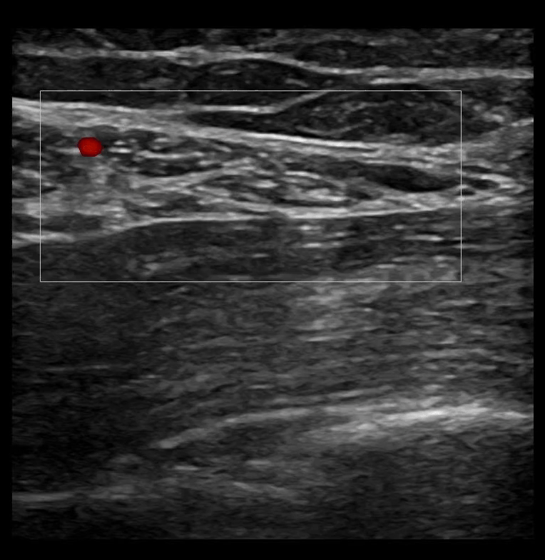
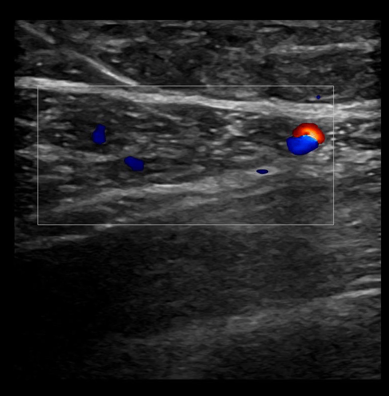
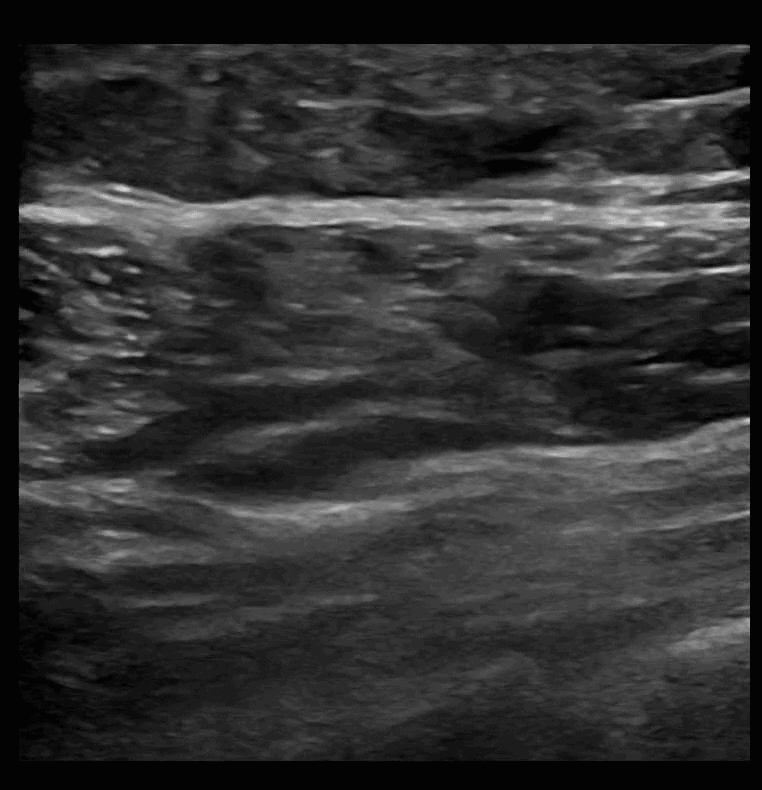
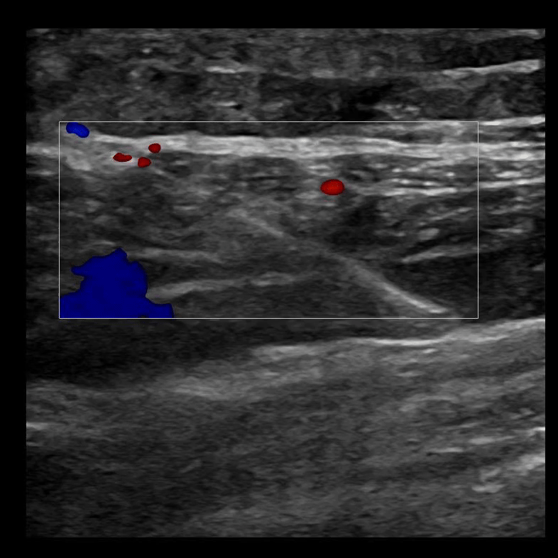
SAPB 2: The latissimus dorsi muscle sits superficial to the serratus anterior muscle. A color box on the latissimus dorsi muscle identifies vasculature.
SAPB 3: Needle is guided in long axis from posterior to anterior with the target being the fascial plane that separates the latissimus dorsi muscle from the serratus anterior muscle.
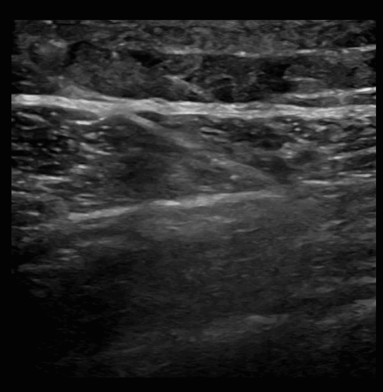
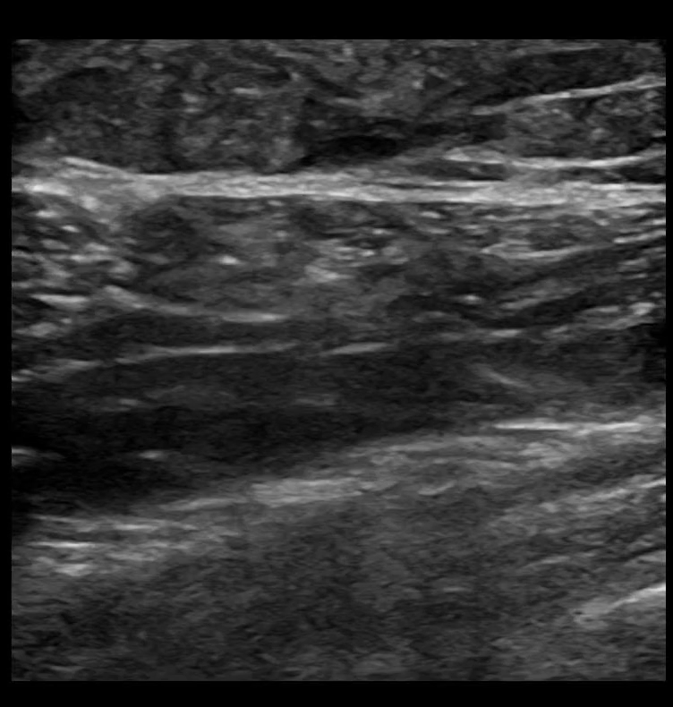
SAPB 4: Unzipping of the latissimus dorsi and serratus anterior muscles with hydrodissection.
SAPB 5: Anesthetic is deposited in the fascial plane between the latissimus dorsi muscle (superficial) and the serratus anterior muscle (deep).
SAPB 6: The ability to follow your needle tip in long axis is an absolute necessity, because deep to the serratus anterior muscle lies rib and pleura.
SAPB 7: The serratus anterior plane block deposits anesthetic in the plane between the latissimus dorsi muscle superficial to it and the serratus anterior muscle deep to it. If done correctly, the two muscles will unzip, or seperate, from each other.
SAPB 8: Needle in long axis being removed. Visualization of needle tip is maintained throughout the entire procedure.