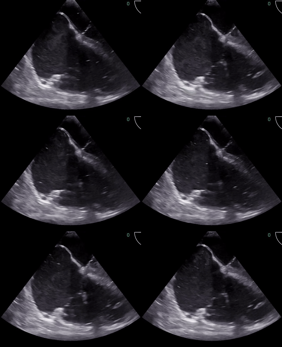
Clip 1: Resuscitative TEE mid esophageal 4 chamber showing massively dilated right atrium and right ventricle.
Image 2: Clot burden retrieved via mechanical thrombectomy.
Adult 70s year old patient presented with syncope. Hemodynamics; BP 125/75, HR 108, T 98.1, resp 18, Sp02 94%, Alert and responsive, ECG showed sinus tach at 106 bpm with a right bundle branch block. POCUS TTE showed right ventricular strain. Immediately anticoagulated based on very high suspicion for PE, CT confirmed suspicion showing saddle PE. Hemodynamics began to deteriorate and clot lysis attempted, but without effect. Progressively losing mental status, the patient was intubated. POCUS TEE mid esophageal 4 chamber view (Clip 1) showed right ventricular failure. Patient was taken to the cath lab for rescue thrombectomy retrieving a massive clot burden (image 2) with improvement of hemodynamics.
The evidence: TEE reduces compression pauses during CPR (Fair et al)

