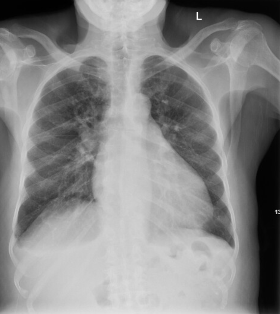
Image 1: Chest radiograph; no pulmonary edema. Please do get your CXR, but as above it will miss pulmonary edema almost 40% of the time.
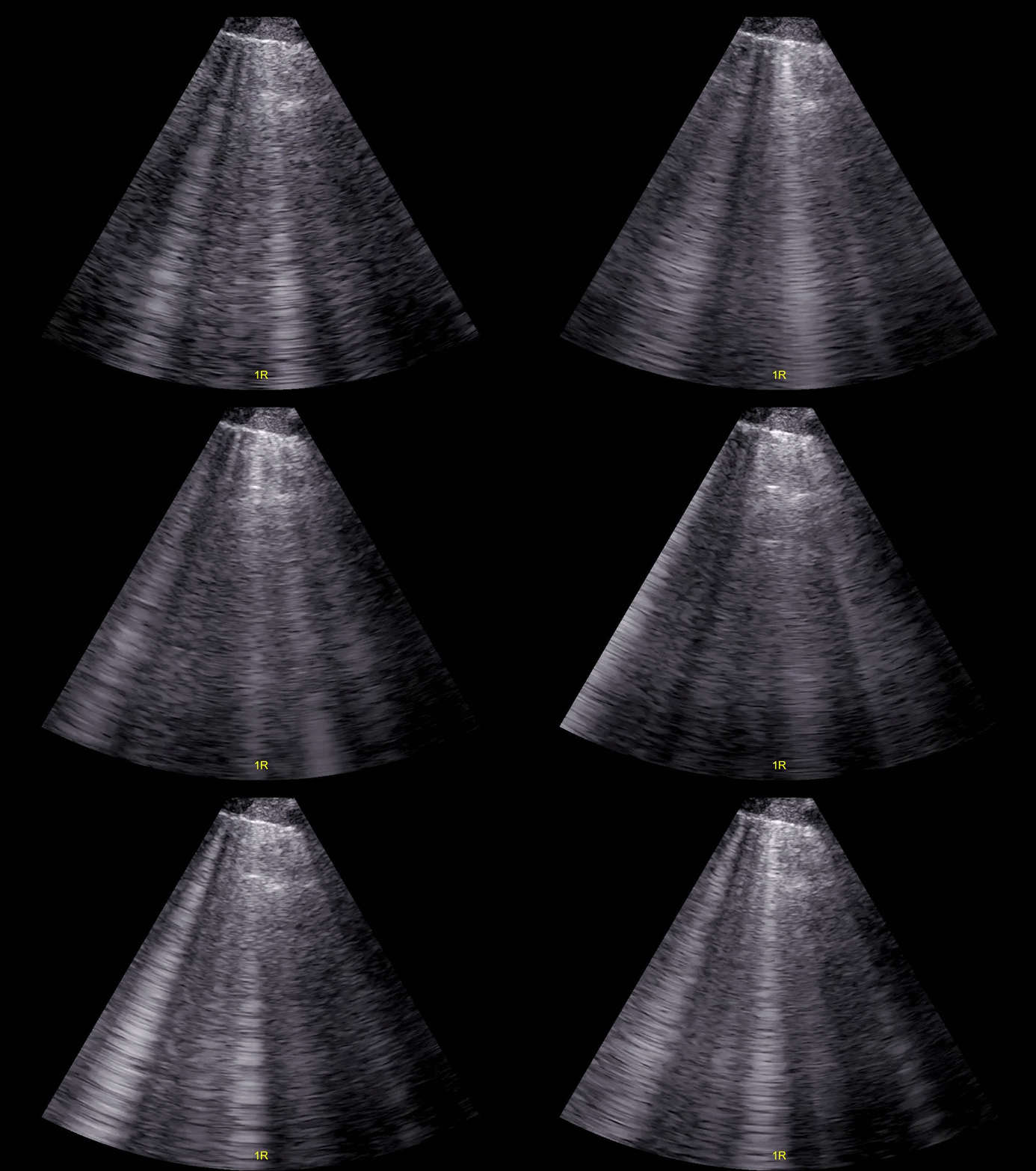
Image 2: POCUS Lung: Right anterior chest: diffuse B lines.
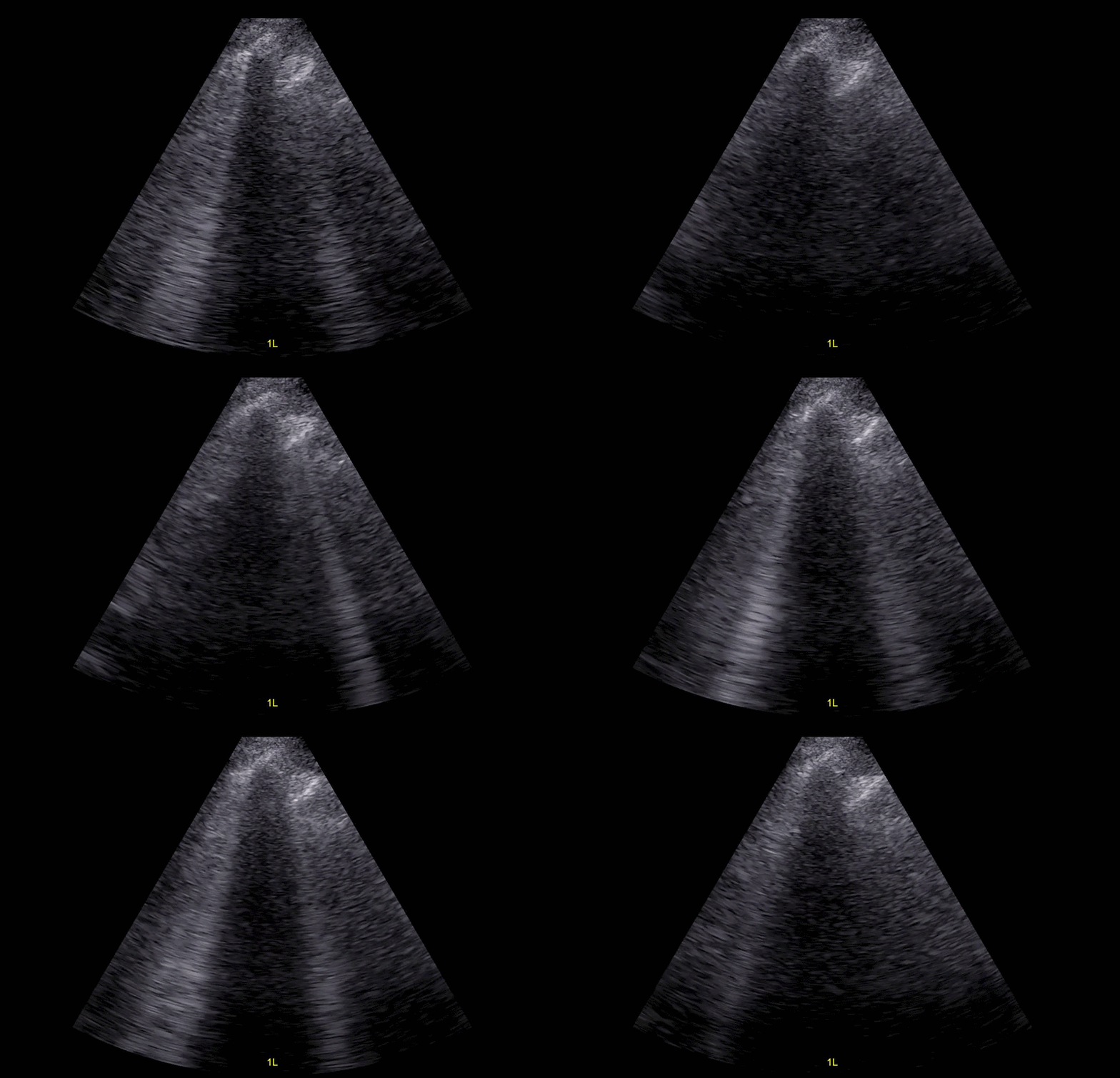
Image 3: Lung POCUS: Left anterior chest also with B lines.
A middle aged patient presents with new onset dyspnea. Let’s keep it simple. With regards to the evaluation of acute heart failure in the emergency department, thanks to Martindale et al, we’ve known for over a decade that:
Radiographic pulmonary congestion is absent 39% of the time (Image 1 above).
BNPs have good negative predictive value.
ECGs do little to nail down the diagnosis of AHF.
Lung ultrasound: B lines have good positive likelihood ratio (Image 2 and 3), and no B lines have a good negative likelihood ratio.
If you’re not already doing so, please add on lung POCUS, it’s your best test for the diagnosis of acute heart failure in the emergency department.